

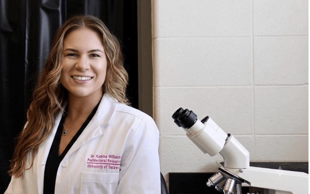
UD study in rats shows specialized imaging can detect hormone-driven brain changes, laying groundwork for future menopause research
Fluctuations in estrogen influence the brain, helping reshape connections between neurons. In a new study using female rats, University of Delaware researchers showed that a specialized imaging technique called magnetic resonance elastography (MRE) can detect estrogen-dependent mechanical changes in the hippocampus, a brain region involved in memory and learning, across the reproductive cycle.
The findings, published in Brain Communications, lay the groundwork for future studies exploring how hormonal changes, including long-term transitions like menopause, affect brain health in women.
“Across the reproductive cycle, the brain adapts, relying more heavily on different regions at different times. This was step one: can we track these changes with MRE in rats, and then eventually in humans?” said first author Katrina Milbocker, a postdoctoral researcher in Curtis Johnson’s laboratory and a UD alumna with a doctorate in behavioral neuroscience.
“Imaging is often viewed as a tool for detecting disease, but it can also help us understand brain health dynamically,” said Johnson, an associate professor of biomedical engineering. “Our ultimate goal is to understand how brain mechanics change across life stages and what those changes can tell us about health and aging.”
Understanding the hormone-hippocampus link
Rodent studies in the 1990s showed that estrogen helps reshape connections between neurons in the hippocampus across the reproductive cycle. The UD researchers suspected those changes might also alter the tissue’s mechanical properties, making it stiffer or softer.
To find out, they turned to MRE, a specialized form of magnetic resonance imaging (MRI). While conventional MRI reveals anatomy and function, MRE measures how the brain acts mechanically, like a material when squeezed, revealing properties such as stiffness and viscosity.
The researchers worked with female rats, which undergo a four-day reproductive cycle called the estrous cycle.
“Rats don’t menstruate, but their estrous cycle has similar hormone fluctuations to humans, allowing us to watch these changes unfold relatively quickly,” Milbocker said.
By scanning the same animals repeatedly across the estrous cycle, the researchers tracked how the hippocampus changed mechanically as hormone levels shifted. As estrogen levels fell between phases of the cycle, tissue viscosity increased, rising by nearly 40% when estrogen production dropped across ovulation.
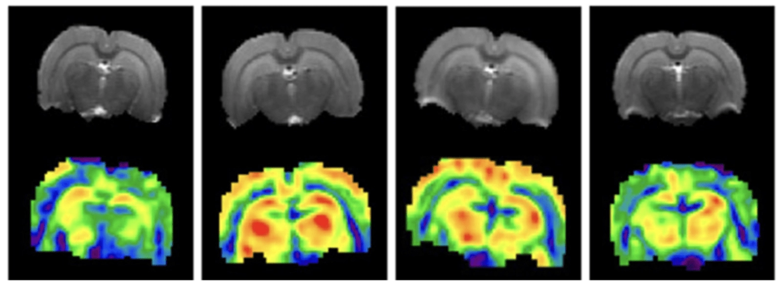
- MRE scans of the rat brain collected at four stages of the reproductive cycle, with color maps showing changes in brain tissue stiffness over the course of the cycle.
To determine whether estrogen was driving the changes, researchers gave the same rats a drug that blocks estrogen receptors. The treatment eliminated the cycle of mechanical changes, suggesting that estrogen signaling was directly responsible for the effects measured by MRE.
After imaging, the researchers collected brain tissue samples to identify the cellular changes behind the MRE measurements. They found shifts in the populations of neurons and astrocytes, cells that help maintain brain function and form interconnected networks that contribute to the brain’s mechanical properties. They also found that estrogen signaling drove the mechanical changes, with the strongest effects seen in astrocytes, which are known to respond to mechanical forces and tissue stiffness. These cells may help link hormone fluctuations to the tissue-level changes detected by MRE, and ultimately to brain function.
Toward clinical translation
While the study links hormone fluctuations to changes in hippocampal mechanics, the question of whether those changes influence brain function remains. A key next step is connecting MRE measurements with cognition, including memory and learning.
Johnson’s team is also extending the work into rat models of menopause, a life stage in which the body produces much lower levels of estrogen. “We suspect there may be a mechanical ‘stuck state’ when estrogen is first depleted, contributing to disrupted cognitive function like brain fog, and we are trying to test this next,” he said.
At the same time, the team is working to translate the findings to humans through parallel rodent and human studies.
“One unique advantage at UD is that the human and animal imaging facilities are in the same building, which makes collaboration easier,” Milbocker said.
Ultimately, they hope to make MRE fast enough to be incorporated into routine MRI exams, ideally adding less than a minute to a standard scan.
“Transitions such as menopause span years, and we want to understand how brain health changes across them and how to support individuals during those periods,” Johnson said.
Other UD co-authors of the work are Tyler Williams, who received his doctorate in biomedical engineering in 2025; doctoral student Sabrina Vander Wiele; undergraduate alumna Emma Zarate; Elise Corbin, assistant professor of biomedical engineering; and Anna Klintsova, professor and chair of the Department of Psychological and Brain Sciences. Funding for the development of methods used in this project was provided by the National Institutes of Health.